Course Content
Module 1 – Step 1 Analysing the rhythm
The heart's electrical activity must originate somewhere, and rhythm provides critical information about its origin, timing, and frequency. But what exactly is rhythm, and how do we describe it? This module explores these questions and more. You will practice analysing rhythm strips, describing rhythms on an ECG, and understanding the significance of the various elements observed when interpreting rhythm.
0/10
Module 2 – Step 2 Describing the p wave
The p wave represents atrial depolarisation. What information can be gleaned from observing the p wave and what approach should one use when describing it? In this module you will practise how to describe the P wave and how to interpret your findings by looking at some examples.
0/8
Module 3 – Step 3 PR interval
The PR interval essentially describes how the electrical impulse is conducted between atria and ventricles. What is a normal PR interval and what does an abnormal interval suggest? In this module you will learn to describe the PR interval and the PR segment using a systematic approach so that you can successfully answer these questions.
0/12
Module 4 – Step 4 Assessing the QRS complex
The QRS complex represents ventricular depolarisation. By examining the QRS complex, we can identify potential structural or conduction abnormalities within the ventricles. In this module, you will learn how to describe and analyse the QRS complex to detect and understand these abnormalities.
0/7
Module 5 – Step 5 ST segment and T wave
The ST segment and T wave relate to ventricular repolarisation. Similar to the QRS complex, analysing the ST segment and T wave can help identify potential structural or conduction abnormalities within the ventricles. In this module, you will learn how to describe this portion of the ECG trace and recognise common pathologies associated with abnormal ST segments and T waves, such as cardiac ischaemia and infarction.
0/14
ECG Mastery Course
About Lesson

CARDIAC DOMINANCE

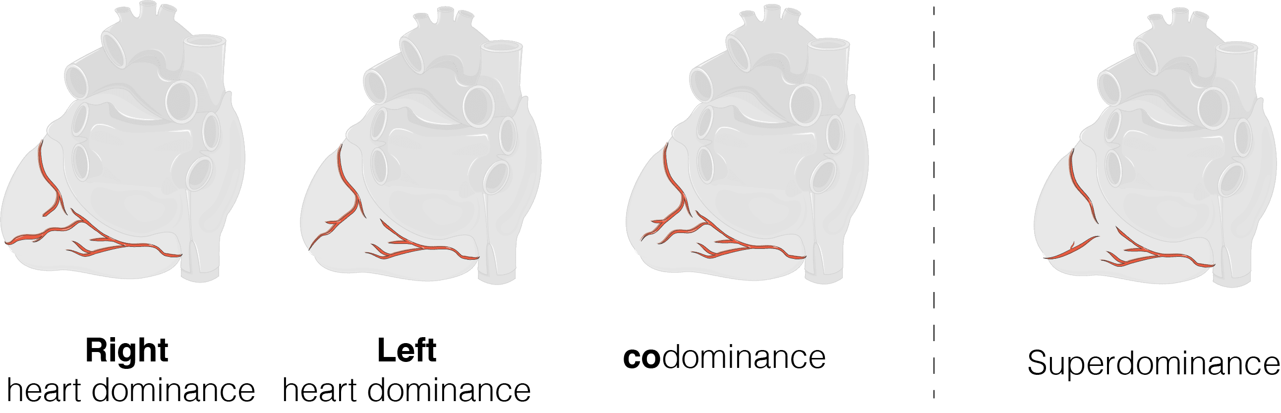
Cardiac dominance refers to which coronary artery supplies the posterior descending artery (PDA). The PDA supplies the inferior and posterior wall of the heart, the posterior portion of the interventricular septum and it typically supplies the AV node. There are largely 3 variations of cardiac dominance (A-C). These include:

  1. Right heart dominance (most common with a prevalence of 80-85%): Origin of the PDA is from the right coronary artery (RCA)
  2. Left heart dominance:  Origin of the PDA is from the left circumflex artery (LCx)
  3. Codominance: The PDA is supplied by both the right coronary artery (RCA) and the left circumflex artery (LCx)
  4. “Super-dominance” is a rare 4th variant where the left anterior descending artery supplies the PDA.

 
CLINICAL APPLICATION

Occlusion of the PDA may cause the following: inferior +- posterior infarction and decreased perfusion to the AV node which may present with an AV block (also known as a heart block).


REFERENCES

  • Shahoud JS, Ambalavanan M, Tivakaran VS. Cardiac Dominance. [Updated 2022 Sep 26]. In: StatPearls [Internet]. Treasure Island (FL): StatPearls Publishing; 2023 Jan-. Available from: https://www.ncbi.nlm.nih.gov/books/NBK537207/
  • Images modified using Servier Medical Art (https://smart.servier.com/) licensed under a Creative Commons Attribution 3.0 unported license (https://creativecommons.org/licenses/by/3.0/)
0% Complete